" width="137.999998613302px"><path d="M 12.985 0 L 0.634 0 L 0 1.472 L 3.601 1.472 L 0.702 8.206 L 5.854 8.206 L 8.753 1.472 L 12.351 1.472 Z M 22.945 0 L 21.566 3.208 L 18.586 3.208 L 19.968 0 L 14.816 0 L 11.283 8.206 L 16.435 8.206 L 17.952 4.679 L 20.93 4.679 L 19.415 8.206 L 24.566 8.206 L 28.097 0 Z M 36.963 0 L 30.261 0 L 26.728 8.206 L 33.433 8.206 C 38.243 8.206 39.925 7.832 41.087 5.135 L 41.971 3.083 C 43.132 0.386 41.776 0 36.966 0 Z M 37.195 2.199 L 35.587 5.94 C 35.324 6.552 34.996 6.734 33.88 6.734 L 32.513 6.734 L 34.806 1.407 L 36.173 1.407 C 37.291 1.407 37.461 1.589 37.198 2.202 Z M 62.002 9.379 C 62.002 9.043 61.997 8.701 61.989 8.35 C 61.979 8.007 61.959 7.71 61.926 7.469 L 61.898 7.272 L 62.239 7.272 L 62.292 7.339 C 62.534 7.646 62.784 7.947 63.042 8.243 C 63.302 8.542 63.615 8.898 63.979 9.307 L 71.686 17.722 L 74.348 17.722 L 74.348 3.604 L 71.575 3.604 L 71.575 12.542 C 71.575 12.915 71.593 13.261 71.626 13.578 L 71.646 13.769 L 71.307 13.769 L 71.254 13.695 C 71.05 13.408 70.815 13.109 70.56 12.806 C 70.3 12.499 69.972 12.128 69.573 11.69 L 62.151 3.604 L 59.201 3.604 L 59.201 17.722 L 61.999 17.722 L 61.999 9.379 Z M 80.02 16.2 C 81.53 17.358 83.535 17.943 85.982 17.943 C 87.257 17.943 88.429 17.762 89.467 17.403 C 90.5 17.047 91.409 16.541 92.172 15.899 C 92.947 15.259 93.545 14.484 93.949 13.59 C 94.354 12.694 94.558 11.715 94.558 10.676 C 94.558 9.693 94.384 8.764 94.038 7.91 C 93.692 7.06 93.162 6.306 92.454 5.668 C 91.692 4.971 90.757 4.425 89.674 4.047 C 88.588 3.668 87.346 3.477 85.982 3.477 C 84.707 3.477 83.535 3.661 82.5 4.027 C 81.467 4.391 80.558 4.901 79.793 5.544 C 79.017 6.211 78.416 7.001 78.002 7.887 C 77.588 8.774 77.381 9.757 77.381 10.811 C 77.381 11.922 77.608 12.95 78.053 13.864 C 78.497 14.778 79.159 15.565 80.017 16.205 Z M 80.755 8.467 C 81.032 7.812 81.421 7.242 81.914 6.774 C 82.409 6.306 83.007 5.945 83.697 5.703 C 84.381 5.464 85.141 5.342 85.957 5.342 C 86.773 5.342 87.56 5.471 88.245 5.728 C 88.934 5.984 89.528 6.348 90.013 6.809 C 90.497 7.269 90.879 7.84 91.146 8.505 C 91.414 9.167 91.55 9.912 91.55 10.721 C 91.55 11.531 91.409 12.32 91.134 12.982 C 90.856 13.645 90.467 14.22 89.975 14.688 C 89.482 15.157 88.886 15.518 88.207 15.759 C 87.53 15.998 86.773 16.12 85.959 16.12 C 85.146 16.12 84.384 15.998 83.699 15.759 C 83.01 15.518 82.411 15.159 81.916 14.688 C 81.424 14.22 81.032 13.647 80.757 12.982 C 80.482 12.323 80.341 11.56 80.341 10.721 C 80.341 9.882 80.482 9.125 80.757 8.47 Z M 106.538 15.831 L 100.392 15.831 L 100.392 3.604 L 97.593 3.604 L 97.593 17.724 L 106.538 17.724 Z M 108.844 3.604 L 111.642 3.604 L 111.642 17.722 L 108.844 17.722 Z M 118.283 11.488 L 123.801 11.488 L 123.781 9.64 L 118.283 9.64 L 118.283 5.474 L 123.912 5.474 L 124.637 3.604 L 115.485 3.604 L 115.485 17.724 L 118.283 17.724 Z" fill="rgb(255, 255, 255)" height="17.943357155935303px" id="ZBDhiyPsr" transform="translate(0 0)" width="124.63716419302727px"/><path d="M 84.808 0 L 84.154 1.87 L 88.656 1.87 L 88.656 16.168 L 0 16.168 L 0 18.417 L 91.477 18.417 L 91.477 17.293 L 91.482 17.293 L 91.482 1.87 L 95.904 1.87 L 96.54 0 Z" fill="rgb(255, 255, 255)" height="18.416533825033483px" id="RINPrdGQR" transform="translate(41.46 3.583)" width="96.54045347780396px"/><path d="M 4.298 10.562 L 10.647 10.562 L 12.339 14.185 L 15.308 14.185 L 8.573 0 L 6.508 0 L 0 14.185 L 2.71 14.185 Z M 7.538 2.847 L 7.743 3.357 L 7.765 3.402 C 7.765 3.402 7.773 3.429 7.778 3.442 L 9.892 8.736 L 5.192 8.736 L 7.543 2.847 Z" fill="rgb(255, 255, 255)" height="14.185338291736393px" id="k8ba4_okS" transform="translate(41.541 3.611)" width="15.30848907851231px"/></g></svg>)
A mini-invasive system for hemorrhoids. In every grade.
THE PROTOCOL
The motor guide
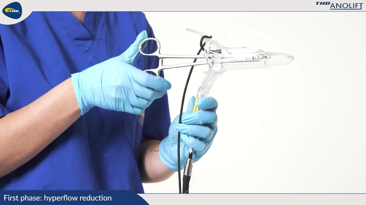
The Doppler locates the artery. The anoscope guides the needle.
One procedure, dual steps
The same anoscope guides the ligation and the mucopexy.
Item by item
Up to six arterial branches ligated, averaging three mucopexy sites.

INSTRUMENTS
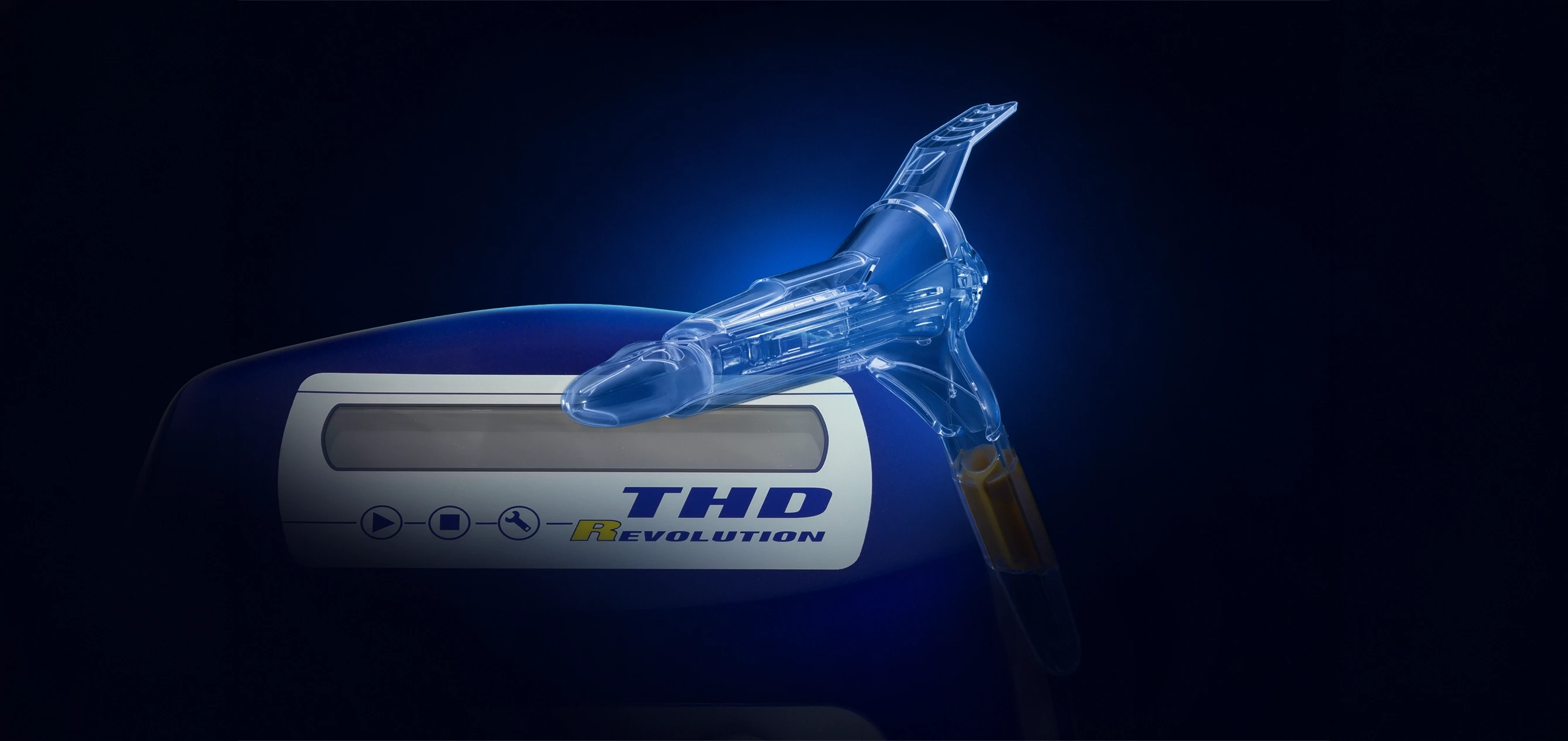
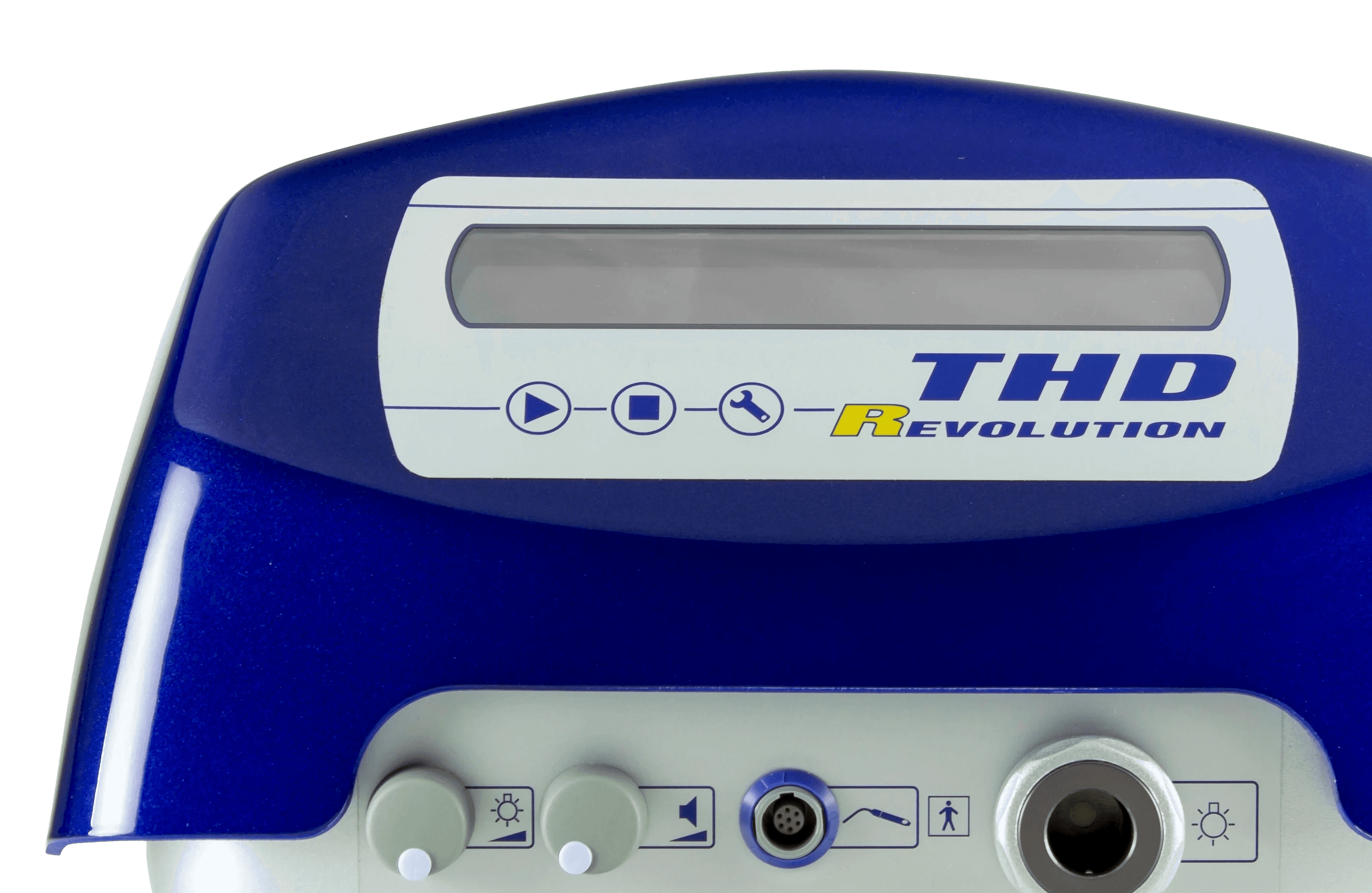
THD Revolution
Doppler console & light emitter. A clear signal locates the ligation site.
INSTRUMENTS
THD Slide
Elliptical atraumatic anoscope. Slide-gate access port.

SUTURING
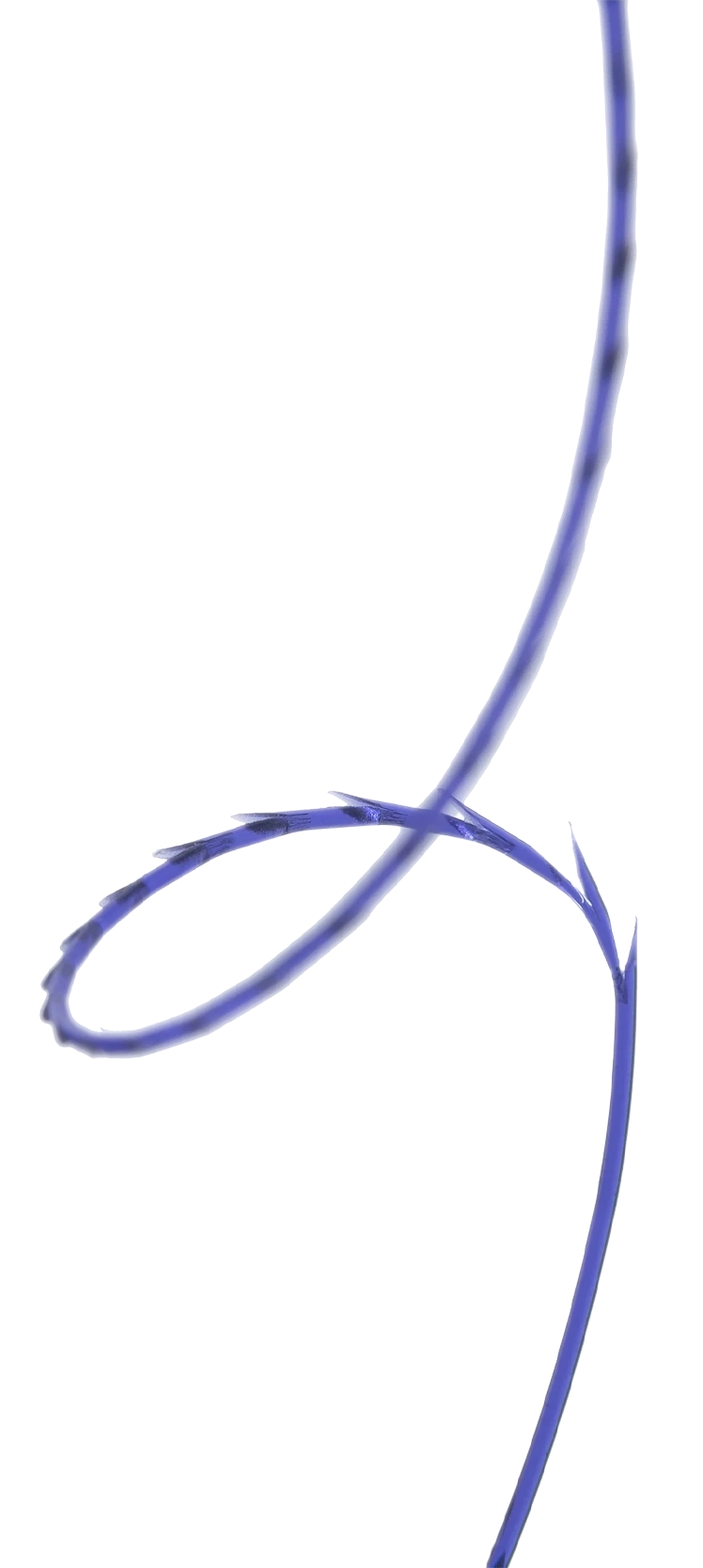
THD Block
Constant tension
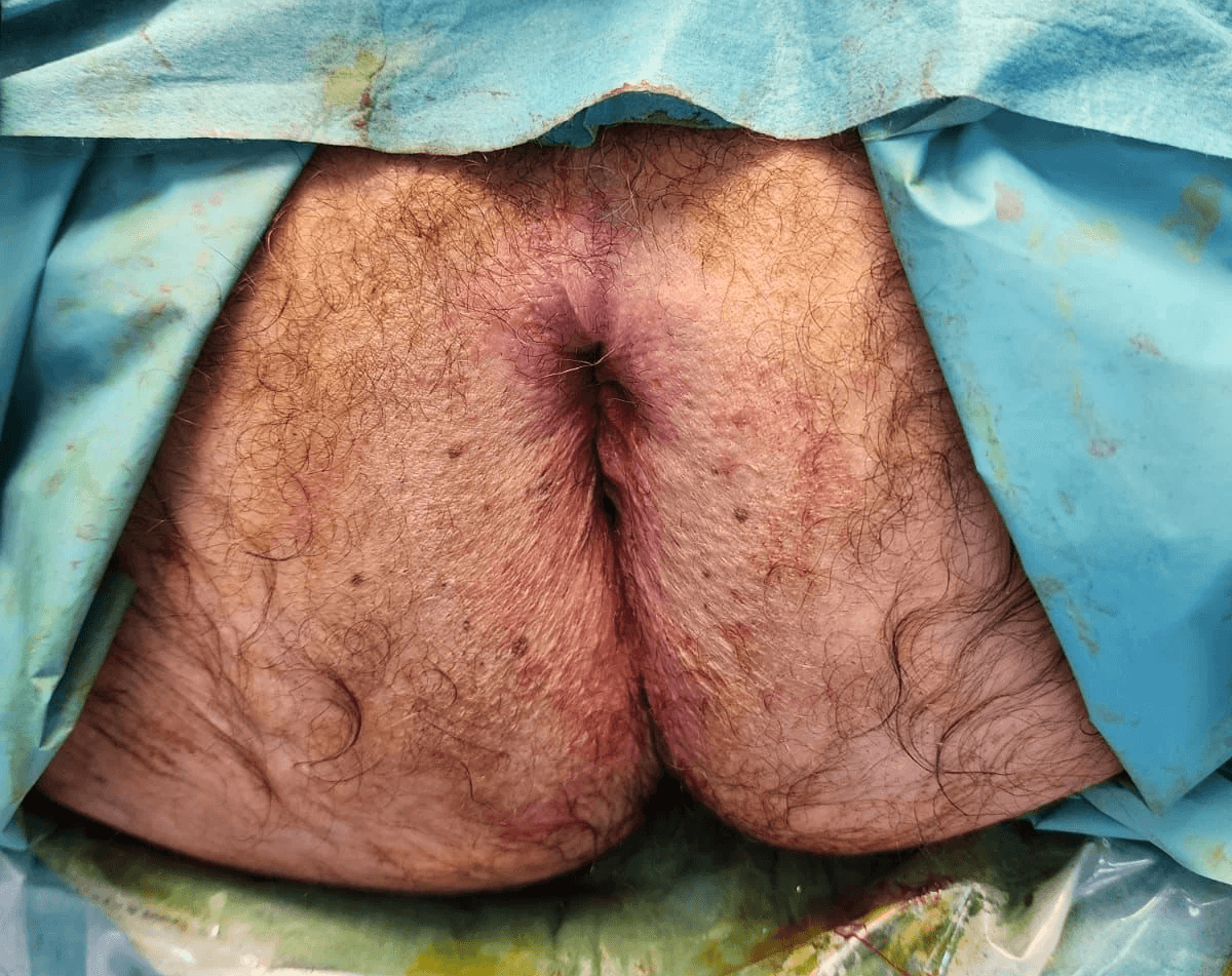
Minimally invasive, non-excisional approach
No tissue excision or energy system; only absorbable suture. Direct visualization procedure with precise artery selection. Excellent outcomes, low complications, and rapid recovery.
Rapid and direct
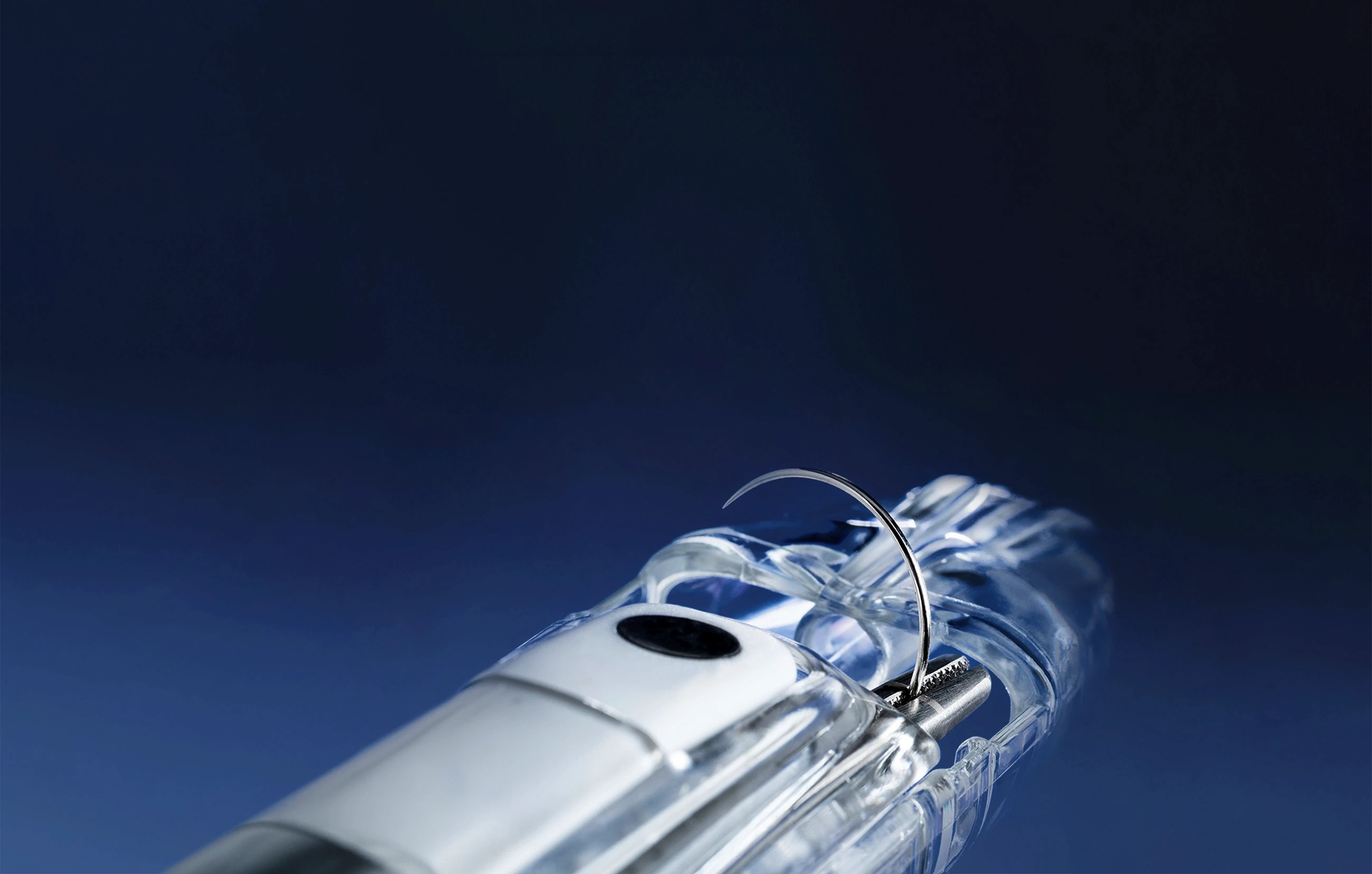
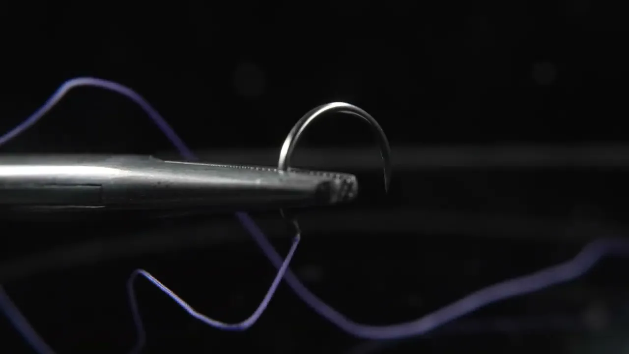
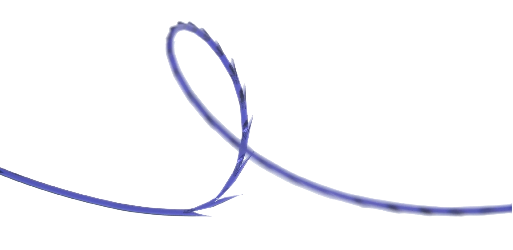
Ligation, combined with THD Anolift, treats all grades of hemorrhoidal disease. THD Block sutures for mucopexy are short, self-locking, and barbed, requiring no knotting and permitting easier management.
Restore patient’s anatomy
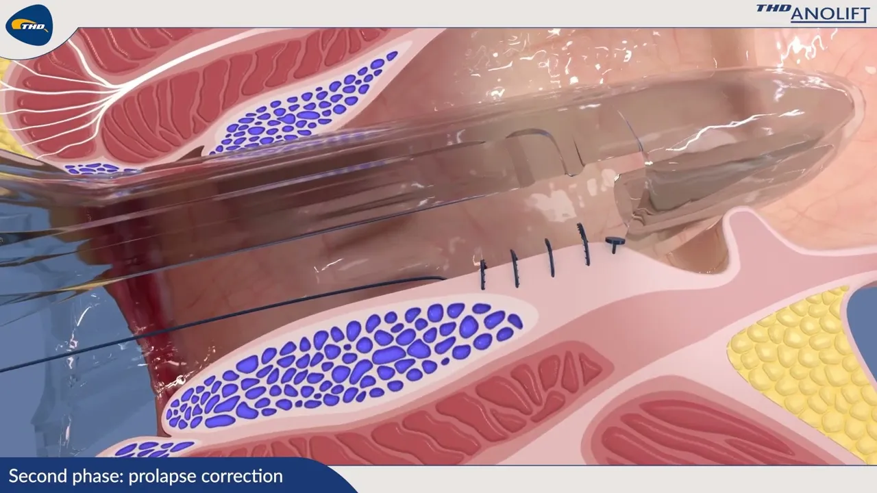
THD Anolift restores the vascular component by reducing flow in superior rectal artery branches via ligation, and restores anatomy by lifting prolapsed tissues during mucopexy.
STEP 1
Ligation

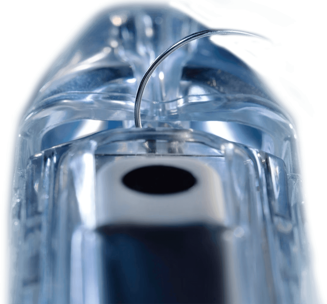
Doppler guided
Precise ligation of the superior rectal artery branches, guided by Doppler
THE PROOF
The ligation acts on vessels. The data confirms it.
In hemorrhoidal patients, mean arterial inflow is significantly elevated
STAGE 2
Mucopexy

Anolift®
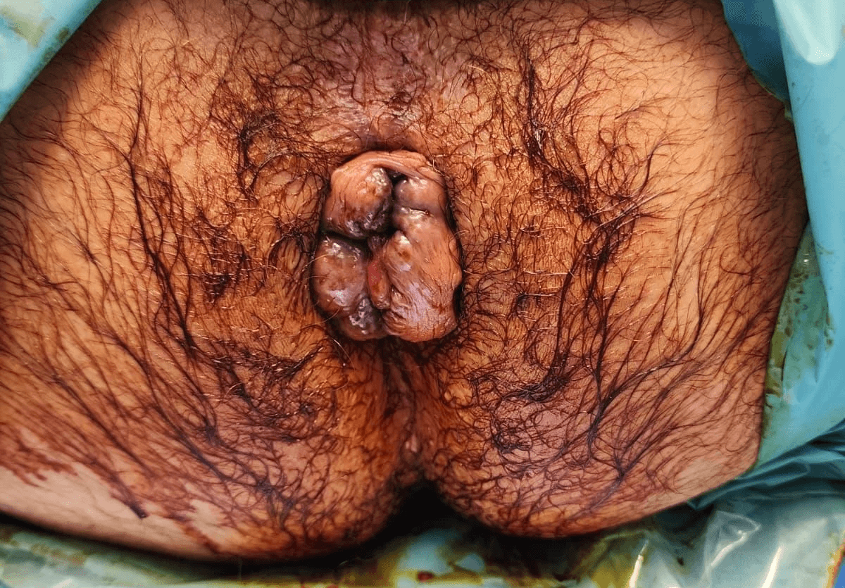
Repositioning of prolapsed tissues, under direct vision
ETIOLOGY
Prolapse origin site
Arterial pressure and connective tissue decay: two factors, one prolapse

Connective tissues
Vessels
Submucosal fibers
9-year-old child

Connective tissues
Vessels
Submucosal connective tissue laxity
70-year-old male
COMPARISON